Overview
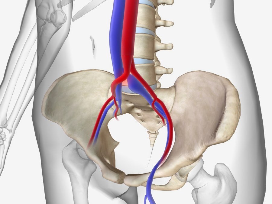
May-Thurner syndrome, also called left iliac vein compression, is a condition in which the pelvic vein draining the left leg is compressed by the main artery running to the right leg. Because the left iliac vein passes through a tight space between the right iliac artery and the spine, it becomes narrowed or completely flattened. The constant pulsation of the artery against the vein can also thicken the vein wall, narrowing it further. When the narrowing becomes severe, it can lead to a blood clot, or deep vein thrombosis (DVT). When there is no associated clot, the condition is called a non-thrombotic iliac vein lesion, or NIVL.
Because the narrowing restricts blood flow out of the lower body, blood pools and creates pressure in the pelvis, leading to a cascade of symptoms that are often unexpected. The classic ones are left leg swelling and a risk of DVT, but emerging data show that iliac vein compression is frequently an unrecognised cause of other chronic problems. The engorged collateral veins that form to bypass the blockage can press directly on nerves, or stretch surrounding veins, producing chronic, heavy pelvic pain.
This pelvic venous pooling is also increasingly linked to postural orthostatic tachycardia syndrome, or POTS, a condition causing dizziness, brain fog, and a rapid heart rate on standing. Because the compressed vein prevents adequate blood from returning to the heart, it can trigger or worsen these autonomic symptoms. Patients with venous compression frequently experience gastrointestinal symptoms that mimic irritable bowel syndrome (IBS) as well, such as severe bloating, constipation, or diarrhea.
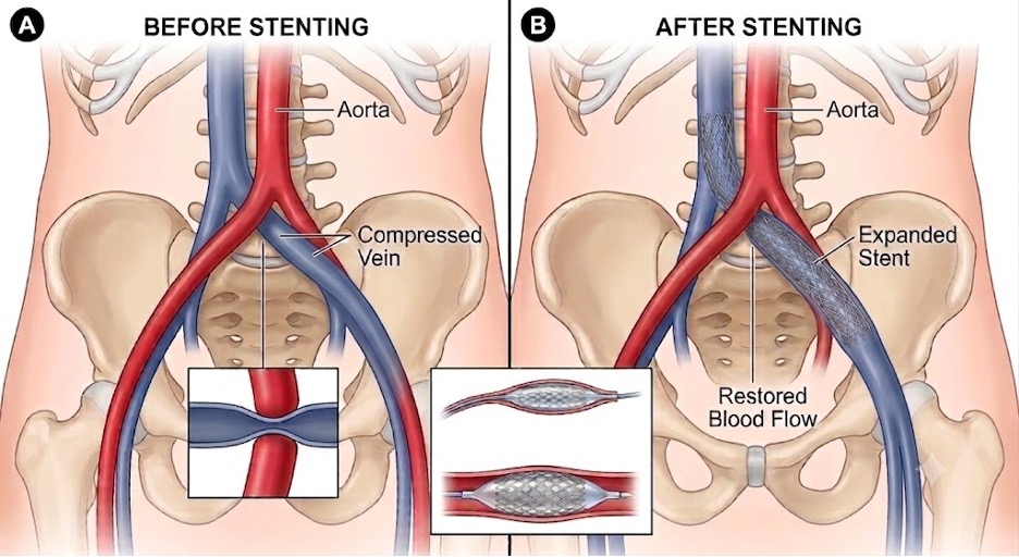
The most effective treatment is a minimally invasive procedure called venoplasty and stenting. Instead of open surgery to reroute the blood vessels, an interventional radiologist works from inside the vein to gently stretch it open and place a permanent support structure that keeps the artery from pinching it closed again.
The procedure safely relieves the pressure in the pelvis and leg, lowers the risk of blood clots, and can significantly improve secondary symptoms such as pelvic pain, POTS, and IBS, with no large surgical incisions and a rapid recovery.
Who it’s for
Most patients considering this procedure have experienced unexplained pelvic pain, swelling, or a feeling of heaviness prominently in the left leg. Many do not discover they have May-Thurner syndrome until they are treated for a deep vein thrombosis in that leg.
A specialised ultrasound of the pelvis, together with a CT scan or MRI, is used to confirm the diagnosis. The imaging will show exactly where the right iliac artery is compressing the left iliac vein.
Who is not a candidate
If your pelvic pain or leg swelling is caused by a different problem, placing a stent will not help. Establishing that the compression is the true source of your symptoms is the purpose of the consultation and the imaging that precedes it.
How the procedure works
1. Accessing the vein
An interventional radiologist cleans and numbs a small area at the groin. A tiny tube called a catheter, about the width of a piece of spaghetti, is inserted through a small needle puncture access into the vein.
2. Navigation and imaging with IVUS
Using real-time X-ray guidance (fluoroscopy), and often an ultrasound probe placed inside the vein itself (intravascular ultrasound, or IVUS), the catheter is navigated to the exact point where the vein is being pinched. You will not feel this happening.
3. Angioplasty: opening the vein
A small deflated balloon is advanced to the narrowed segment and briefly inflated, stretching the vein open and pushing the compressing artery out of the way.
4. Stent placement
To prevent the vein from collapsing again, a stent, a flexible metal mesh tube, is placed. It expands to fit the vein wall precisely and acts as a permanent scaffold, keeping blood flowing freely. The catheter is then removed and a small bandage applied.
What to expect on the day
When you arrive, our team will get you comfortable and start an IV. You will be given “twilight” sedation, so you will be relaxed and sleepy but breathing on your own, without a breathing tube or general anesthesia.
The procedure itself typically takes 45 minutes to an hour. Afterward you will rest in our recovery room for about an hour while the sedation wears off and our team monitors your leg. Because there are no major incisions, you go home the same day.
Recovery
Recovery is brief. On day one, plan to rest at home. You may have mild soreness at the groin access site and an ache in your lower back or pelvis as your body adjusts to the newly placed stent. This is easily managed with pain relievers.
By the end of the first week most patients feel back to normal. You can typically return to a desk job or light duties within two to three days. We ask that you avoid heavy lifting and strenuous exercise for 7 to 10 days so the access site heals properly.
What is normal, and when to call
Mild bruising at the entry site and a temporary ache in your back are completely normal. Call our office immediately if you experience sudden or severe leg swelling, worsening pain that medication does not relieve, or active bleeding at the access point.
Risks and considerations
Stenting for May-Thurner syndrome is a highly safe, minimally invasive procedure, but as with any procedure there are potential risks.
Common, minor side effects include bruising at the access site and a temporary backache.
Rare but more serious risks include infection, injury to the blood vessel, the stent shifting out of position, or a blood clot forming inside the stent. Because the procedure is performed by a highly trained specialist using advanced imaging, the risk of complications is very low, typically under 2%. If an issue does arise, our team is fully equipped to manage it safely.
Results and evidence
The clinical evidence supporting stenting for May-Thurner syndrome is robust. Studies show a technical success rate of over 95%, meaning the vein is successfully opened and stented.
For patients with swelling and pain, the large majority see a dramatic improvement in symptoms within weeks. For patients who have had a DVT, stenting significantly reduces the risk of another clot forming in the future.
The results are durable. Modern stents are designed to last a lifetime, and it is very rare for a stented vein to collapse again.
Common questions
- Is it painful?
- The procedure is well tolerated. We numb the entry site with local anesthesia and provide IV sedation to keep you relaxed and sleepy, and you will not feel the catheter moving inside your body. You may feel a brief moment of pressure in your lower back when the balloon is inflated. Afterward, a mild backache for a few days is common and easily managed with pain medicine.
- Will insurance cover it?
- Yes. Because May-Thurner syndrome is a diagnosed medical condition that restricts blood flow and increases the risk of dangerous blood clots, the procedure is generally covered by Medicare and most major insurance carriers. Our office will work with your insurance provider to verify your exact benefits before proceeding.
- How soon will I notice a difference?
- Many patients notice that the pelvis and leg feel lighter and less swollen within the first few days after the procedure. It can take a few weeks for the residual swelling to resolve completely as your body adjusts to the improved blood flow.
- What happens if it doesn’t work?
- In the rare event that symptoms return, we will perform an ultrasound to check the stent. Occasionally scar tissue builds up inside it, narrowing the vein again. If that happens, a very similar minor balloon procedure can be done to stretch it back open.