Overview
A vascular malformation is a tangle of blood vessels that formed incorrectly before birth. Unlike a birthmark or a haemangioma, it does not go away on its own. It grows with the person, and it often becomes symptomatic in adolescence or adulthood, or after an injury, a hormonal change, or a pregnancy.
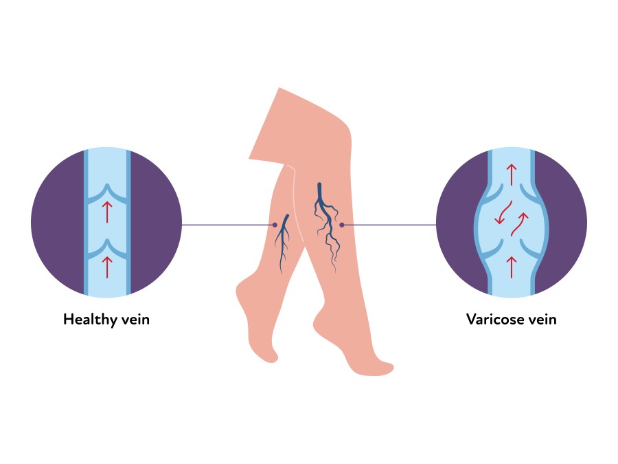
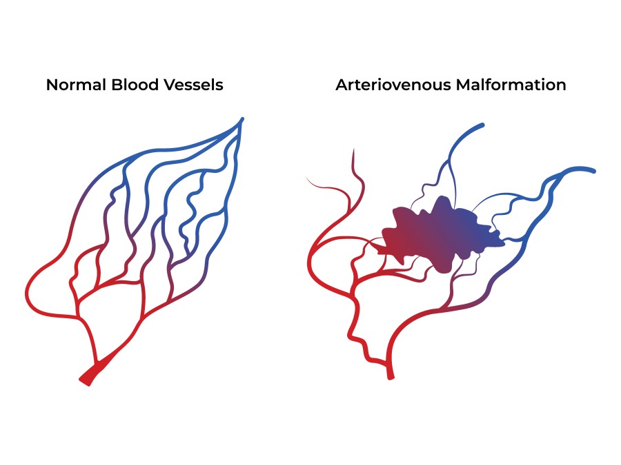
They are grouped by the vessels involved and by how fast blood moves through them. Slow-flow malformations include venous malformations, which are soft, bluish, and typically ache and swell when the limb is dependent or after use, and lymphatic malformations, which swell and can become infected. Fast-flow malformations are arteriovenous malformations (AVMs), where arteries connect directly to veins without the capillary bed in between, producing warmth, a pulsation, pain, and sometimes bleeding or skin breakdown.
Embolization and sclerotherapy treat these lesions from the inside. A catheter or needle is guided into the abnormal vessels under imaging, and an agent is delivered that closes them off permanently. The surrounding normal tissue is left alone, which is what makes the treatment precise in a way surgery around these lesions rarely can be.
The goal is control of symptoms: less pain, less swelling, less bleeding, and better function and appearance, without open surgery and with a recovery measured in days rather than weeks.
Who it’s for
Most patients come to the clinic because a malformation has become painful, has grown, has begun to interfere with function, or is causing bleeding or repeated infections. Many have been told for years that nothing could be done, or have been offered surgery that carried a high chance of recurrence.
Diagnosis is made with imaging rather than biopsy. Ultrasound with Doppler shows whether the flow is fast or slow, and MRI shows the true extent of the lesion, which is almost always greater than what is visible from the outside. For fast-flow lesions, a diagnostic angiogram maps the feeding arteries, the connection point, and the draining veins, and that map determines the whole treatment plan.
Getting the classification right matters more here than in almost any other area of this practice. Malformations are frequently misdiagnosed, and the treatment for a venous malformation is not the treatment for an AVM.
Who is not a candidate
Small, painless malformations that are not growing may simply be watched, with treatment held in reserve. Where a lesion involves critical structures, treatment may be limited to the symptomatic portion rather than the whole. And a haemangioma of infancy is a different condition altogether, often resolving on its own or responding to medication, so establishing the diagnosis first is essential.
How the procedure works
1. Mapping the lesion
Treatment begins with imaging, and for fast-flow lesions with a diagnostic angiogram. We need to know exactly where the abnormal connection sits, what feeds it, and what drains it. Treating the wrong part of an AVM, such as blocking only the feeding arteries, makes the lesion recruit new supply and become harder to treat later.
2. Reaching the abnormal vessels
For slow-flow venous and lymphatic malformations, a fine needle is placed directly into the lesion through the skin under ultrasound guidance. For fast-flow AVMs, a catheter is passed through a small needle puncture access in the artery, usually at the top of the thigh, and a microcatheter is navigated into the heart of the lesion.
3. Sclerotherapy or embolization
For venous and lymphatic malformations, a sclerosant is injected. It irritates the lining of the abnormal channels so they scar down and close. For arteriovenous malformations, an embolic agent such as a liquid glue, a liquid embolic polymer, or coils is delivered precisely into the connection point to close it permanently.
4. Confirming the result
Imaging at the end of the procedure confirms that the treated portion has closed and that the normal vessels around it are flowing as they should. The needle or catheter is removed and a simple dressing is applied.
Treatment is usually staged
This is the most important thing to understand about vascular malformations. Except in small lesions, treatment is delivered in several sessions spaced weeks to months apart, each targeting a portion of the lesion. That is deliberate: it is safer, it allows the tissue to settle between sessions, and it produces a better long-term result than attempting everything at once.
What to expect on the day
When you arrive, our team will get you comfortable and start an IV. Most sessions are done with sedation, and some, particularly extensive sclerotherapy or treatment in a sensitive area, are done under general anesthesia. We will tell you which applies to your session well before the day.
The session itself typically takes one to two hours, depending on the size and location of the lesion. You will rest in our recovery room for about an hour afterward.
Most patients go home the same day. An overnight stay is occasionally arranged when the lesion is large, when significant swelling is expected, or when it involves the airway or another sensitive area.
Recovery
Expect the treated area to swell, and expect it to look worse before it looks better. Swelling and firmness typically peak in the first two to three days and then subside over one to two weeks. This is the intended inflammatory response, not a complication. Ice, elevation, and over-the-counter pain relievers manage it well, and for larger lesions we may prescribe a short course of steroids to limit the swelling.
Most patients return to desk work and light activity within two to three days, and to full activity within one to two weeks depending on the site treated. If a limb was treated, compression may be recommended.
The lasting result develops over months as the treated channels scar down and contract. You will be seen in follow-up, and further sessions, if planned, are usually scheduled six to twelve weeks apart.
What is normal, and when to call
Swelling, firmness, bruising, and discomfort in the treated area are all expected. Call our office if you develop skin blistering or breakdown over the lesion, spreading redness, a fever above 101°F, new numbness or weakness, dark urine, or pain that is escalating rather than settling.
Risks and considerations
These procedures are performed in specialist centres for good reason. They are safe in experienced hands, but they carry more specific risks than the other treatments described on this site, and we would rather set them out plainly.
Expected effects include swelling, bruising, firmness, and discomfort in the treated area for one to two weeks.
The main specific risks are injury to the overlying skin, which can blister or ulcerate if the sclerosant reaches it; injury to a nearby nerve, causing numbness or weakness that is usually temporary; infection; and, with larger sclerotherapy volumes, effects on the blood or kidneys that we monitor for and manage. For fast-flow AVMs there is the additional risk of the embolic agent passing beyond the intended target.
These risks are reduced by treating in stages, by using real-time imaging throughout, and by matching the agent and the volume to the lesion rather than to a protocol. Our team is equipped to manage complications immediately if they arise.
Results and evidence
Embolization and sclerotherapy are the accepted first-line treatment for most symptomatic vascular malformations, and have largely replaced surgery as the primary approach.
For slow-flow venous and lymphatic malformations, the published series report meaningful improvement in pain, swelling, and size in the large majority of patients, typically after more than one session. Lymphatic malformations, particularly the larger-cyst type, tend to respond especially well.
For arteriovenous malformations the honest picture is more nuanced. A true cure is achievable in some lesions, particularly small, well-defined ones with a single connection point, but many are managed rather than cured: the aim is durable control of pain, bleeding, and growth. Incompletely treated AVMs can recruit new blood supply over time, which is why complete closure of the connection point, rather than the feeding arteries, is the technical goal.
Across all types, the realistic measure of success is symptom control and function rather than disappearance on a scan, and long-term follow-up is part of the treatment rather than an optional extra.
Common questions
- Is it painful?
- The procedure itself is done with sedation or general anesthesia, so you will not feel it. Afterward, the treated area is typically sore, swollen, and firm for one to two weeks, more so than with the other procedures described on this site. That discomfort is expected, it is a sign the treatment is working, and it is managed with ice, elevation, and pain medication.
- How many sessions will I need?
- It depends on the size and type of the lesion. A small venous malformation may need one or two. A large or extensive one commonly needs three to six sessions, spaced six to twelve weeks apart. We will give you an honest estimate after the imaging, and we will reassess as we go.
- Will insurance cover it?
- Generally yes. When a vascular malformation causes pain, swelling, bleeding, repeated infection, or functional impairment, treatment is medically necessary rather than cosmetic, and is covered by most carriers. Our office will verify your benefits before you proceed.
- Can it come back?
- Treated channels stay closed, but a malformation is a congenital condition and untreated portions can enlarge over time, particularly with hormonal changes or after an injury. Arteriovenous malformations in particular can recruit new supply if the connection point was not fully closed. This is why follow-up matters, and why recurrence is usually treated as another stage rather than as a failure.
- Would surgery be better?
- Rarely, as a first step. Malformations do not have clean edges, so surgical removal is often incomplete, and an incompletely removed lesion tends to recur and can be harder to treat afterward. Surgery has a role, usually after embolization has reduced the lesion and the bleeding risk, or for removing residual bulk once the vessels are closed.